
ASA Abstract Reviews
Ultrasound Image of Expansion of the Epidural Space in Pediatric Epidural Analgesia
Masahiro Yagihara, MD; Aki Uemura, MD, PhD; Yoshiki Nakajima, MD, PhD
Hamamatsu University School of Medicine, Hamamatsu, Japan
(Summarized and submitted by Masahiro Yagihara, MD)
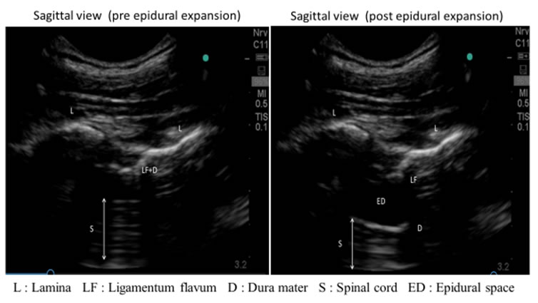
Epidural analgesia is playing important role not only with anesthesia but also postoperative period. Since epidural analgesia is accomplished using surface landmarks and blind needle insertion, the epidural approach is frequently administered for adult patients but less for pediatrics. In our hospital, we ensure pediatric epidural procedures safely with using a customized special kit for pediatric epidurals, the micro drip infusion methods, after confirming the distance from the skin to the lumbar epidural space before needle insertion by the formula previously reported(1,2). Besides, to confirm the expansion of the epidural space in pediatrics by real-time ultrasound imaging may improve efficacy and safety in comparison with the blind epidural needle insertion. Therefore we evaluated spread of the epidural space by ultrasound technique in 40 pediatric patients(mean age 1.6±1.8 year, mean body weight 8.7±4.9 kg) retrospectively.
We recognized lamina, ligementum flavum, dura mater, and spinal cord with micro convex probe (Sono Site M-turbo C11x/8-5 Fuji Film Japan) by sagittal plain. The visibility rate of dura mater was 82.5%. Epidural space was expanded from 0.92±0.33mm to 5.1±2.5mm by direct injection of fluid through Touhy needle. The epidural space was significantly expanded immediately after reaching the needle to the epidural space in micro drip infusion technique. The real-time ultrasound image of the needle insertion to the epidural space might indicate the technique is suitable for not only confirmation of epidural space expansion but pre-scanning. In conclusion, ultrasound images are useful for observing expanding the epidural space in pediatric epidural analgesia. We thought it was necessary to find relationship between the age, bodyweight, local anesthetic spread and the expansion of epidural space prospectively.
References
- M Yamashita et al. Identification of the epidural space with an i.v. micro-drip infusion set in infants and children. Can J Anaesth 1990 May;37(4-2)S99
- A Uemura et al. A formula for determining the distance from the skin to the lumbar epidural space in infants and children. Pedi Anaesth 1992 Dec;2(4):305-7
![]()